
For Physical Therapy & Veterinary Rehabilitation Professionals

EVIDENCE • EXPERIENCE • PRACTICAL APPLICATION

Clinical Insights‍ ‍

Connect Knowledge to Practice

Start with Clinical Insights to understand the diagnosis, identify key rehabilitation priorities, and connect directly to the protocols, courses, and GoGoCharlie® templates that support clinical decision-making and patient care.

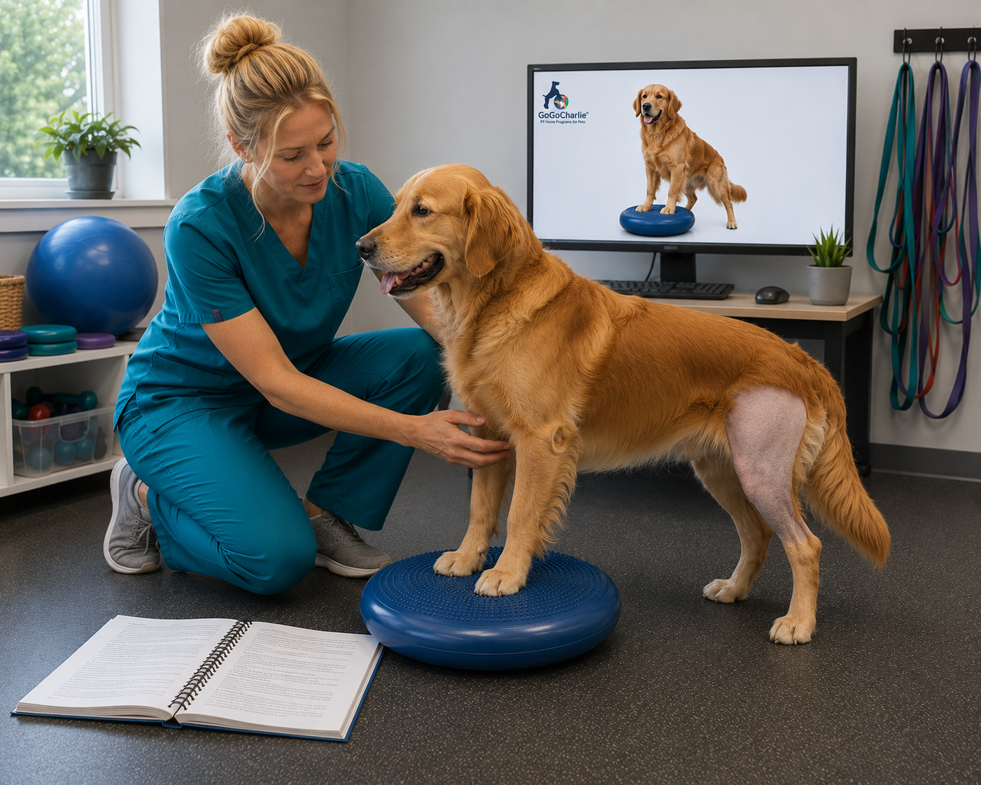
Woman in scrubs working with dog
Download icon
Video icon
GoGoCharlie silhouette

Explore Protocols
Immediate access to the linked workbook.

View Protocols →

Watch the Clinical Course
Access diagnosis-specific rehabilitation videos.

Start Learning →

Try GoGoCharlie® Free
Create client ready HEPs with diagnosis templates.

Create Programs →

Every Clinical Insight connects clinical education, treatment planning, and home program delivery — one pathway from learning to patient care.

Muscle, Orthopedics, Neurology, Oncology Sasha A Foster Muscle, Orthopedics, Neurology, Oncology Sasha A Foster

Medial Shoulder Instability Rehabilitation

Medial Shoulder Instability Rehabilitation Clinical Insights

Overview

Medial shoulder instability (MSI) is a common cause of forelimb lameness in active and sporting dogs. Injury to the medial glenohumeral ligament, joint capsule, and subscapularis tendon results in shoulder laxity, pain, and reduced athletic performance. Successful rehabilitation emphasizes protecting healing tissues while progressively restoring dynamic shoulder stability.

Key Clinical Points

Diagnosis requires more than an abduction angle

  • Shoulder abduction testing is helpful but should never be used alone.

  • Clinical examination and advanced imaging provide a more accurate diagnosis.

Conservative treatment is the first choice

  • Most Grade I and II injuries respond well to rehabilitation.

  • Surgery is generally reserved for severe or non-responsive cases.

Dynamic stability is the goal

  • Rehabilitation focuses on restoring muscular control rather than simply reducing pain.

  • Progressive loading helps protect the healing ligament while improving shoulder function.

 
Successful medial shoulder instability rehabilitation restores shoulder stability through controlled loading, progressive strengthening, and protection of healing tissues.
 

Rehabilitation Priorities

  • Protect injured medial shoulder structures.

  • Control pain and inflammation.

  • Restore dynamic shoulder stability.

  • Progress strengthening without increasing instability.

  • Return to pain-free functional activity.

Evidence Snapshot

  • Conservative rehabilitation is recommended as first-line treatment for most dogs with MSI.

  • Clinical rehabilitation programs report excellent return to function in appropriately managed cases.

  • There is currently no evidence that one surgical technique consistently produces superior outcomes.

  • Successful outcomes depend on matching treatment progression to tissue healing and shoulder stability.

Clinical Pearls

  • Treat instability before progressing strengthening.

  • Grade the injury to guide rehabilitation progression.

  • Monitor the entire kinetic chain for compensatory dysfunction.

  • Return to sport should be based on function—not time alone.


Continue Learning

View the complete rehabilitation protocol.

Deliver home exercise programs and client education.

Watch clinical demonstrations, treatment techniques, and practical applications.


Medial Shoulder Instability Rehabilitation FAQ

Read More
Muscle, Orthopedics, Neurology, Oncology Sasha A Foster Muscle, Orthopedics, Neurology, Oncology Sasha A Foster

Elbow Dysplasia Rehabilitation

Elbow Dysplasia Rehabilitation Clinical Insights

Overview

Elbow dysplasia is one of the most common developmental orthopedic diseases affecting the canine elbow. It includes fragmented medial coronoid process (FMCP), osteochondritis dissecans (OCD), ununited anconeal process (UAP), and elbow incongruity. These conditions alter normal joint loading, leading to pain, reduced range of motion, lameness, and progressive osteoarthritis. Successful rehabilitation combines medical management, weight optimization, progressive therapeutic exercise, and long-term load management to improve function and quality of life.

Key Clinical Points

Radiographs do not tell the whole story

  • Imaging findings often do not correlate with the dog's functional limitations.

  • Rehabilitation progression should be based on clinical function, gait quality, and response to loading—not imaging alone.

Weight management is one of the most effective treatments

  • Maintaining a lean body condition significantly decreases joint loading.

  • Obesity accelerates osteoarthritis progression and worsens long-term outcomes.

Rehabilitation is central to long-term success

  • Controlled strengthening, underwater treadmill therapy, and gradual loading improve mobility and reduce compensation.

  • Lifelong management is often necessary, even after surgery.

 
Successful elbow dysplasia rehabilitation focuses on restoring comfortable function through progressive loading, weight management, and lifelong protection of the joint.
 

Rehabilitation Priorities

  • Reduce pain and inflammation while protecting the medial elbow compartment.

  • Restore comfortable elbow range of motion and symmetrical limb loading.

  • Strengthen the shoulder, elbow, and entire kinetic chain using progressive closed-chain exercise.

  • Optimize body condition to reduce mechanical stress on the joint.

  • Monitor compensatory movement patterns and adjust activity based on clinical response rather than radiographs.

Evidence Snapshot

  • Recent systematic reviews suggest that selected dogs with fragmented medial coronoid disease may achieve outcomes comparable to surgery when managed conservatively with structured rehabilitation.

  • Long-term studies demonstrate that radiographic severity does not reliably predict clinical progression, emphasizing functional assessment during rehabilitation.

  • Underwater treadmill therapy has been shown to improve elbow range of motion and stride length while reducing joint loading.

  • Maintaining a lean body weight remains one of the strongest evidence-based interventions for slowing osteoarthritis progression.

Clinical Pearls

  • Evaluate the entire kinetic chain—shoulder, cervical spine, trunk, and opposite limb frequently develop compensatory dysfunction.

  • Dogs often continue exercising despite pain; owners should regulate activity rather than allowing self-limitation.

  • Progress exercise based on recovery after activity, gait symmetry, and comfort—not predetermined timelines.

  • Even following successful surgery, long-term rehabilitation and weight management remain essential to minimize osteoarthritis progression.


Continue Learning

View the complete rehabilitation protocol.

Deliver home exercise programs and client education.

Watch clinical demonstrations, treatment techniques, and practical applications.


Carpus Hyperextension Rehabilitation FAQ

To learn more, download the Elbow Dysplasia Treatment Protocol Workbook today.

Read More
Muscle, Orthopedics, Neurology, Oncology Sasha A Foster Muscle, Orthopedics, Neurology, Oncology Sasha A Foster

Biceps Brachii Injury Rehabilitation

Biceps Brachii Rehabilitation Clinical Insights

Overview

Biceps brachii injuries are a common cause of chronic front limb lameness in dogs. These injuries may involve the muscle, tendon, surrounding tissues, or occur with other shoulder conditions. Successful rehabilitation focuses on identifying the source of pain, protecting healing tissues, rebuilding strength, and safely returning the patient to activity.

Key Clinical Points

Accurate diagnosis guides treatment
○ Identifying the biceps brachii as the source of pain is essential for selecting the right treatment approach.
○ Imaging can help determine injury severity and guide rehabilitation decisions.

Tendon healing requires gradual progression
○ Improved movement does not always mean the tendon has fully healed.
○ Exercises should progress based on comfort, strength, and tissue tolerance.

The whole shoulder must be considered
○ Biceps brachii injuries often occur with other shoulder conditions.
○ Addressing strength, stability, and movement patterns supports long-term recovery.

 
Successful tendon rehabilitation requires balancing protection of healing tissues with progressive loading to restore strength and function.
 

Rehabilitation Priorities

• Reduce pain and inflammation while protecting irritated biceps tissue.
• Maintain comfortable shoulder mobility without overstressing healing structures.
• Restore neuromuscular control and shoulder stabilization.
• Progressively rebuild tendon load tolerance and muscular strength.
• Monitor for recurrence, compensation, or signs that additional diagnostics are needed.

Evidence Snapshot

• Musculoskeletal ultrasound is an important diagnostic tool for evaluating biceps tendon fiber changes, tendon enlargement, mineralization, and response to treatment.

• Conservative management has shifted toward structured rehabilitation with functional reassessment rather than relying on fixed recovery timelines.

• Progressive exercise loading is a key component of tendon rehabilitation, with early low-load activation progressing toward strengthening as tissue tolerance improves.

Clinical Pearls

• Evaluate the entire shoulder, not only the biceps tendon; compensatory changes and concurrent pathology may influence recovery.

• Avoid progressing activity based only on improved gait — tendon remodeling and functional strength require additional time.

• Flare-ups should be treated as a clinical signal to reassess loading, activity level, and contributing factors.


Continue Learning

View the complete rehabilitation protocol.

Deliver home exercise programs and client education.

Watch clinical demonstrations, treatment techniques, and practical applications.


Carpus Hyperextension Rehabilitation FAQ

To learn more, download the Biceps Brachii Injury Treatment Protocol Workbook today.

Read More
Muscle, Orthopedics, Neurology, Oncology Sasha A Foster Muscle, Orthopedics, Neurology, Oncology Sasha A Foster

Carpus Arthrodesis Rehabilitation

Carpus Arthrodesis Rehabilitation Clinical Insights

Overview

Carpus arthrodesis is performed to restore stability following traumatic or chronic carpal collapse by surgically eliminating motion at the affected joint(s) and achieving surgical stabilization. Rehabilitation after partial or pancarpal arthrodesis focuses on protecting the surgical construct, managing distal limb edema, preserving mobility of adjacent joints, and restoring functional strength. Because loss of carpal motion alters thoracic limb biomechanics, rehabilitation emphasizes compensatory mobility, proximal limb strengthening, controlled loading, and safe progression based on clinical assessment and radiographic evidence of healing.

Key Clinical Points

  • Protect the Fusion During Healing

    • Surgical fixation restores stability, but progression must be guided by clinical findings and radiographic confirmation of osseous healing.

    • Consistent weight bearing, controlled swelling, and absence of increasing pain are key indicators for advancement.

  • Treat the Entire Thoracic Limb Kinetic Chain

    • Loss of carpal motion eliminates normal elastic recoil during terminal stance.

    • Rehabilitation should address digit mobility, elbow and shoulder function, and proximal strength to support long-term compensation.

  • Progress Loading Based on Function, Not Time Alone

    • Recovery requires gradual restoration of strength, endurance, and functional mobility while protecting the surgical construct.

    • Changes in lameness, swelling, or comfort should guide modification of activity progression.

 
Successful outcomes depend on maintaining distal limb range of motion to compensate for loss of motion at the carpus.
 

Rehabilitation Priorities

  • Improve functional weight-bearing to promote bone healing.

  • Maintain distal and proximal joint PROM and AROM.

  • Educate caregivers on importance of controlled activity until radiographic evidence of healing.

Evidence Snapshot

  • Stress radiography remains an important diagnostic tool for confirming carpal hyperextension and identifying instability location.

  • Complete rupture of palmar support structures has limited healing capacity under normal weight-bearing forces, making surgical stabilization the preferred approach for many severe injuries.

  • Pancarpal arthrodesis provides predictable stability when multiple carpal levels are affected, while partial arthrodesis may be considered when instability is limited.

Clinical Pearls

  • Monitor the full limb, not only the carpus: Elbow, shoulder, cervical, and thoracolumbar compensation may influence recovery.

  • Functional goals should reflect patient needs, such as comfortable walking, stair navigation, and return to daily activities.


Continue Learning

View the complete rehabilitation protocol.

Deliver home exercise programs and client education.

Watch clinical demonstrations, treatment techniques, and practical applications.


FAQ for Carpus Arthrodesis

To learn more, download the Carpus Arthrodesis Treatment Protocol Workbook today.

Read More
Muscle, Orthopedics, Neurology, Oncology Sasha A Foster Muscle, Orthopedics, Neurology, Oncology Sasha A Foster

Carpus Hyperextension Rehabilitation

Carpus Hyperextension Clinical Insights

Overview

Carpus hyperextension occurs when the palmar support system fails, affecting ligaments, fibrocartilage, and flexor structures. Successful rehabilitation depends on identifying instability severity, protecting healing tissues, and rebuilding dynamic muscular support.

Key Clinical Points

  • Accurate grading drives decision-making

    • Stress radiographs remain the diagnostic cornerstone for identifying instability.

    • Musculoskeletal ultrasound may help characterize palmar ligament, fibrocartilage, and tendon involvement.

  • Stability determines treatment pathway

    • Some injuries may respond to OTC or custom orthotic support combined with rehabilitation.

    • Complete disruption of palmar support structures has limited healing capacity with conservative care alone.

  • Dynamic support matters

    • Carpal and digital flexor strength contribute to functional stabilization.

 
Successful outcomes depend on matching tissue healing, stability needs, and exercise progression to each patient’s presentation.
 

Rehabilitation Priorities

  • Protect injured palmar structures while maintaining safe limb use.

  • Restore controlled terminal stance stability.

  • Improve neuromuscular activation and dynamic muscular support.

  • Address secondary compensations throughout the kinetic chain.

  • Use functional assessment and tissue tolerance to guide progression.

Evidence Snapshot

  • Imaging improves diagnosis and injury classification.

  • Orthotic management may support selected ligament injuries.

  • Progressive loading supports long-term functional recovery.

Clinical Pearls

  • Evaluate the whole patient, not only the carpus; compensatory changes may occur in adjacent joints and the spine.

  • Orthotic success depends on fit, skin tolerance, owner compliance, and integration with rehabilitation.

  • Terminal stance control is a key functional indicator during recovery.


Continue Learning

View the complete rehabilitation protocol.

Deliver home exercise programs and client education.

Watch clinical demonstrations, treatment techniques, and practical applications.


Carpus Hyperextension Rehabilitation FAQ

To learn more, download the Carpal Hyperextension Treatment Protocol Workbook today for complete rehabilitation guidelines and clinical decision-making resources.

Read More